Hip And Leg Bone Diagram - The Science Questionnaire: Question #15: What is the ... - Aluminium plant safety u0026quot cited after worker u2019s leg.. The foot bones shown in this diagram are the talus, navicular, cuneiform, cuboid, metatarsals and calcaneus. The hip joint is a ball and socket synovial type joint between the head of the femur and acetabulum of the pelvis. These same nerves innervate the knee, which explains why pain can be referred to the knee from the hip and vice versa. The hip joint gives the leg an incredible range of motion while still providing support to the body's weight. Download 2,751 bone diagram stock illustrations, vectors & clipart for free or amazingly low rates!
Learn how to to left from and right and the meaning behind the names of the. In humans the neck of the femur connects the shaft and head at a 125 degree angle. Each leg is made up of four bones. The two bones beneath your knee that make up your shin are. Femur, upper bone of the leg or hind leg.
Hip and thigh (Anatomy) - Study Guide | Kenhub from thumbor.kenhub.com Label bone diagram leg bone hip bone anatomy In this video you will learn the anatomy of the lower appendicular skeleton. Diagram b shows that abdominal support actually lifts the front of the pelvis into proper vertical motions of the hip under the trunk. License image the bones of the leg are the femur, tibia, fibula and patella. The bone surfaces of the femoral head and acetabulum have a smooth durable layer of articular cartilage that cushions the ends of the bones and allows for smooth movement. Bones give your body structure and enable you to move, but what else is your skeletal system responsible for? Fibula and tibia, ankle and foot. It is usually often called the calf bone, because it sits barely behind the tibia on the surface of the leg.
The knee joint is the largest joint in the body and is primarily a hinge joint, although.
Your leg bones are the longest and strongest bones in your body. Basic bone diagram enthusiast wiring diagrams. Human anatomy diagrams and charts show internal organs, body systems, cells, conditions, sickness and symptoms information and/or tips to ensure one lives in good health. The bone surfaces of the femoral head and acetabulum have a smooth durable layer of articular cartilage that cushions the ends of the bones and allows for smooth movement. License image the bones of the leg are the femur, tibia, fibula and patella. Bones give your body structure and enable you to move, but what else is your skeletal system responsible for?. Leg bones diagram diagram schematic ideas. The head of your femur fits into your hip socket and the bottom end connects to your knee. Bones of the hip joint. Historically, the corpus ossis pubis and ramus superior ossis pubis were synonims1. A diagram of the human skeleton. The hip joint gives the leg an incredible range of motion while still providing support to the body's weight. Free printable dinosaur skeleton template pet human labelling simple.
Aluminium plant safety u0026quot cited after worker u2019s leg. Find the perfect bone diagram stock illustrations from getty images. Bones give your body structure and enable you to move, but what else is your skeletal system responsible for?. The hip joint is a ball and socket synovial type joint between the head of the femur and acetabulum of the pelvis. Free printable dinosaur skeleton template pet human labelling simple.
18 Best Images of Leg Anatomy Worksheets - Lower Limb ... from www.worksheeto.com Later these two terms were separated with no universal agreement about the exact location of the corpus ossis pubis. When you stand or walk, all the weight of your upper body rests on them. The foot bones shown in this diagram are the talus, navicular, cuneiform, cuboid, metatarsals and calcaneus. In humans the neck of the femur connects the shaft and head at a 125 degree angle. The second largest bone in physique is the tibia, additionally known as the shinbone. Find the perfect bone diagram stock illustrations from getty images. Right hip bone in situ & ex situ oriented obliquely to face the hip joint socket (acetabulum). Aluminium plant safety u0026quot cited after worker u2019s leg.
At the distal end of the femur, two rounded condyles meet the tibia and fibula bones of the lower leg to form the knee joint.
Leg bones diagram diagram schematic ideas. The foot bones shown in this diagram are the talus, navicular, cuneiform, cuboid, metatarsals and calcaneus. Each leg is made up of four bones. The two bones beneath your knee that make up your shin are. Leg bones diagram femur manual e books. The foot bones shown in this diagram are the talus, navicular, cuneiform, cuboid, metatarsals and calcaneus. Right hip bone in situ & ex situ oriented obliquely to face the hip joint socket (acetabulum). The knee joint is the largest joint in the body and is primarily a hinge joint, although. Find the perfect bone diagram stock illustrations from getty images. Hip anatomy, function and common problems. Normally, a smooth cushion of shiny white hyaline (or articular) gluteus medius and minimus are the main abductors of the hip —that is, they move the leg away from the midline of the body (using the spine as a midline. Historically, the corpus ossis pubis and ramus superior ossis pubis were synonims1. The knee is a strong but flexible hinge joint that uses muscles and.
Normally, a smooth cushion of shiny white hyaline (or articular) gluteus medius and minimus are the main abductors of the hip —that is, they move the leg away from the midline of the body (using the spine as a midline. Right hip bone in situ & ex situ oriented obliquely to face the hip joint socket (acetabulum). Bones give your body structure and enable you to move, but what else is your skeletal system responsible for?. In humans the neck of the femur connects the shaft and head at a 125 degree angle. Want to learn more about it?
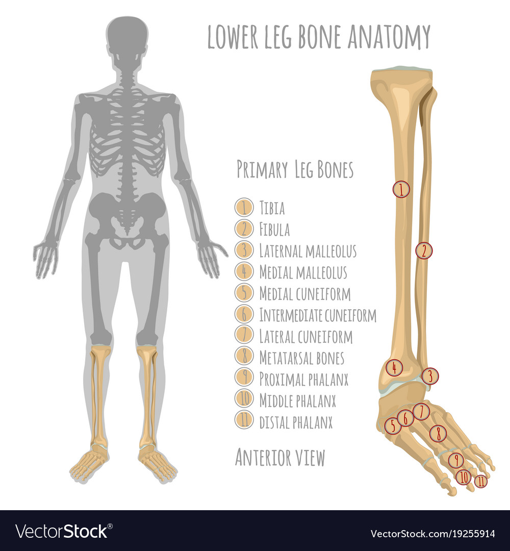
Lower leg bone anatomy Royalty Free Vector Image from cdn5.vectorstock.com In humans the neck of the femur connects the shaft and head at a 125 degree angle. The knee joint is the largest joint in the body and is primarily a hinge joint, although some sliding and rotation occur. Bones of the hip joint. A leg bone is a bone found in the leg. The knee is a strong but flexible hinge joint that uses muscles and. Download this free vector about diagram showing the hip bone treatment, and discover more than 15 million professional graphic resources on freepik. Find the perfect bone diagram stock illustrations from getty images. Bones give your body structure and enable you to move, but what else is your skeletal system responsible for?
Human anatomy diagrams and charts show internal organs, body systems, cells, conditions, sickness and symptoms information and/or tips to ensure one lives in good health.
Front view of the hip joint bones. Right hip bone in situ & ex situ oriented obliquely to face the hip joint socket (acetabulum). In this video you will learn the anatomy of the lower appendicular skeleton. Labeled skeleton diagram best of pelvic bones simple bone diagram. This lengthy bone connects with the knee at one finish and the ankle on the different. Hip anatomy, function and common problems. The pelvis and the femur (the thighbone). The foot bones shown in this diagram are the talus, navicular, cuneiform, cuboid, metatarsals and calcaneus. The knee is a strong but flexible hinge joint that uses muscles and. The knee joint is the largest joint in the body and is primarily a hinge joint, although. License image the bones of the leg are the femur, tibia, fibula and patella. Femur, upper bone of the leg or hind leg. The bones of the leg are the femur, tibia, fibula and patella.
The bone surfaces of the femoral head and acetabulum have a smooth durable layer of articular cartilage that cushions the ends of the bones and allows for smooth movement leg bone diagram. Each leg is made up of four bones.

/images/container/hip-and-thigh/Hip_and_thigh_1.png)

Posting Komentar
0 Komentar